
Va
Aliasing velocity
- Manually set value on the echocardiography machine.
- It´s used to calculate: EROA.
- Nyquist limit: 50-70cm/s
- Baseline is shifted in the direction of mitral regurgitation jet to 30-40cm/s.
- Aliasing occurs: If the blood flow from the probe (blue) exceeds the Va speed (38.5cm/s).


VCW
Vena contracta width
- Narrowest portion of jet as emerges from mitral orifice.
- Vena contracta is diameter of EROA (Effective Regurgitant Orifice Area).
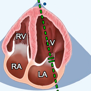
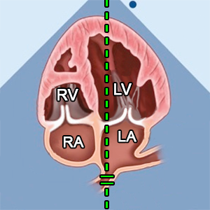
- A4C (Apical 4 chamber) or A2C (Apical 2 chamber)
- Zoom mode (focused on mitral valve)
- Mid-systole (ECG: The beginning of T wave).
- Color doppler
- Nyquist limit (Aliasing velocity): 30-40cm/s


PISAr
Proximal isovelocity surface area radius
- The radius of PISA is measured from the surface of the hemisphere to the narrowest portion of jet (Vena contracta).
- Vena contracta is narrowest portion of jet as emerges from mitral orifice.
- The flow convergence zone is the zone of increased flow velocity before the regurgitant orifice.
- It´s used to calculate: EROA.
- A4C (Apical 4 chamber) or A2C (Apical 2 chamber)
- Zoom mode (focused on mitral valve)
- Mid-systole (ECG: The beginning of T wave).
- Color doppler
- Nyquist limit (Aliasing velocity): 30-40cm/s


Vmax MR
Peak velocity mitral regurgitation
- It´s used to calculate: EROA.
- A4C (Apical 4 chamber)
- Mid-systole (ECG: The beginning of T wave).
- CW doppler
- Place the cursor between mitral leaflet tips.


EROA
Effective Regurgitant Orifice Area
- EROA = 2π x PISAr2 x Va / Vmax MR
- Is the narrowest area of mitral regurgitation flow.


A wavedominant
Dominant mitral A wave (A wave > E wave)
- A4C (Apical 4 chamber).
- Diastole (ECG: The end of T wave - R wave).
- PW doppler.
- Sample volume between mitral leaflet tips.
- Suggest mild mitral regurgitation.


RegJetsoft density
Regurgitant jet soft density
- Density and contour of regurgitant jet
- Density is proportional to the number of red blood cells reflecting the signal
- A4C (Apical 4 chamber).
- Systole (ECG: R wave - The end of T wave).
- CW doppler
- Compare density with nonregurgitant flow
- Dense signal suggests significant MR, whereas a faint signal is likely to be mild or trace MR.


RegJetarea
Regurgitation jet area
- It´s used to calculate: RegJet/LA area
- A4C (Apical 4 chamber)
- Zoom mode (focused on mitral valve)
- Systole (ECG: R wave - The end of T wave).
- Color doppler
- Nyquist limit (Aliasing velocity): 50-70cm/s


LA area
Left atrial area
- It´s used to calculate: RegJet/LA area
- A4C (Apical 4 chamber)
- Zoom mode (focused on mitral valve)
- End-systole (ECG: The end of T wave).


RegJet/LA area
Ratio RegJetarea/LA area
- RegJet/LA area = RegJetarea/LA area x 100
- Ratio area of the jet to the left atrium area.


PVreversal flow
Pulmonary vein systolic flow reversal
- Systolic flow reversal in the pulmonary veins
- A4C (Apical 4 chamber).
- Systole (ECG: R wave - The end of T wave)
- PW doppler
- Sample volume from right (or left) pulmonary vein.
- Color doppler (helps with blood flow identification).
- Systolic flow reversal in the pulmonary veins suggests severe mitral regurgitation.


SVAoV
Stroke volume of aortic valve
- SVAoV = CSALVOT x VTILVOT
- It´s used to calculate: RegVolMV
- Calculate it in the top menu: AV


MVdiameter
Mitral valve diameter
- It´s used to calculate: CSAMV and SVMV
- A4C (Apical 4 chamber)
- Diastole (ECG: The end of T wave - R wave)
- Method: Inner edge to inner edge


VTI MV
Velocity time integral mitral valve inflow
- It´s used to calculate: SVMV
- A4C (Apical 4 chamber)
- Diastole (ECG: The end of T wave - R wave)
- PW doppler
- Sample volume between mitral leaflet tips.
- Trace along the edge of the modal velocity to measure the area under the curve.


SVMV
Stroke volume of mitral valve
- SVMV = CSAMV x VTI MV
- It´s used to calculate: RegVolMV and RFMV


RegVolMV
Regurgitant volume of mitral regurgitation
- RegVolMV = SVMV - SVAoV
- It´s used to calculate: RFMV.


RFMV
Regurgitant fraction of mitral valve
- RFMV = RegVolMV / SVMV x 100
- Regurgitation fraction of mitral regurgitation.


LV EF
Left ventricular ejection fraction (Biplane Method of Discs)
- LV EF = (LVEDV - LVESV) / LVEDV x 100
- Ejection fraction is the predominant method for assessing global systolic function
- and is derived from the LVEDV and LVESV.
- Calculate it in the top menu: Left ventricle


LA volume
Left atrial volume (Biplane)
- A4C (Apical 4 chamber) and A2C (Apical 2 chamber).
- End-systole (ECG: The end of T wave).
- Preferred technique is method of disk summation (modified biplane).
- Trace the LA inner border, excluding the area under the MV annulus, pulmonary veins, and LA appendage from the A4C and A2C views.
- Calculate it in the top menu: Atria and IVC


LVEDV
Left ventricular end-diastole volume
- A4C (zoomed left ventricle), A2C (zoomed left ventricle).
- End-diastole (ECG: R wave).
- Preferred technique is Biplane Method of Discs (modified Simpson’s rule).
- Measured from the A4C and A2C views (preferably an LV focused view) tracing the endocardial – blood pool interface
- Papillary muscles should be excluded from the cavity tracing.
- Maximize LV area and avoid foreshortening.
- Calculate it in the top menu: Left ventricle


MVA
Mitral valve area
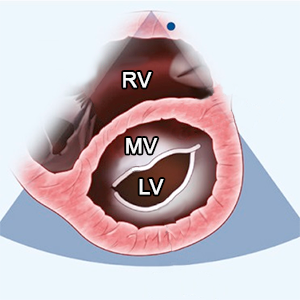
- PSAX (level of MV)
- Mid-diastole (ECG: The beginning of P wave)
- An anatomical orifice is measured.


meanPG MV
Mitral valve mean pressure gradient
- A4C (Apical 4 chamber)
- Diastole (ECG: The end of T wave - R wave)
- CW doppler
- Place the cursor in the middle of mitral valve.
- Trace along the edge of the modal velocity to measure the area under the curve.


RVSP(SPAP)
Right ventricular systolic pressure
- RVSP(SPAP) = 4 x Vmax TR2 + RAP
- RVSP = SPAP (Systolic Pulmonary Artery Pressure)
- RVSP = SPAP (in absence of RVOT obstruction)
- Calculate it in the top menu: RV pressure.
References:
Recommendations for Cardiac Chamber Quantification by Echocardiography in Adults: An Update from the ASE and EACVI (2015)
Recommendations for the Evaluation of LV Diastolic Function by Echocardiography: An Update from the ASE and EACVI (2016)
Recommendations on the use of echocardiography in adult hypertension: a report from the EACVI and the ASE (2015)
Recommendations on the Echocardiographic Assessment of Aortic Valve Stenosis: A Focused Update from the EACVI and the ASE (2017)
ASE Recommendations for Noninvasive Evaluation of Native Valvular Regurgitation (2017)
Guidelines for performing a comprehensive TTE examination in adults: Recommendations from the ASE (2018)
Guidelines for the Echocardiographic Assessment of the Right Heart in Adults: ASE, EACVI, ESC, CSE (2010)
Guidelines for the diagnosis and management of acute pulmonary embolism ESC, ERS (2019)
Echocardiography in aortic diseases: EAE recommendations for clinical practice (2010)
Echocardiographic assessment of valve stenosis: EAE/ASE recommendations for clinical practice (2009)
ESSENTIAL ECHOCARDIOGRAPHY A Companion to Braunwald’s Heart Disease
Coronary Artery Territories (Echocardiography Illustrated Book 4)