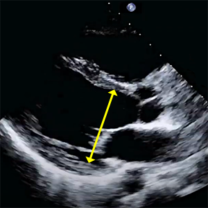

LVIDs
Left ventricular internal dimension at end-systole
- PLAX (Parasternal long axis).
- End-systole (ECG: The end of T wave).
- Measure perpendicular to the long axis of the LV, at or immediately below the level of the mitral valve leaflet tips.


LVIDd
Left ventricular internal dimension at end-diastole
- PLAX (Parasternal long axis).
- End-diastole (ECG: R wave)
- Measure perpendicular to the long axis of the LV, at or immediately below the level of the mitral valve leaflet tips.


IVSd
Interventricular septum thickness at end-diastole
- PLAX (Parasternal long axis).
- End-diastole (ECG: R wave)
- Measure perpendicular to the septum wall.
- Interventricular septum is measured only et end-diastole.


PWd
Left ventricular posterior wall thickness at end-diastole
- PLAX (Parasternal long axis).
- End-diastole (ECG: R wave)
- Measure perpendicular to the posterior wall.
- Posterior wall is measured only et end-diastole.


RWT
Relative wall thickness
- RWT = (2 x PWd) / LVIDd
- PLAX (Parasternal long axis).
- Calculation according to the formula: (2 x PWd) / LVIDd
- Relative Wall Thickness helps calculate if the ventricular morphology has altered.
- RWT reports the relationship between the wall thickness and cavity size.
- RWT allows further classification of LV mass (concentric, eccentric hypertrophy).


LV mass
Left ventricular mass
- LV mass = 0.8 x 1.04 x [(IVSd + LVIDd + PWd)3 - LVIDd3] + 0.6g
- PLAX (Parasternal long axis).
- Left ventricular mass and left ventricular mass indexed to body surface area estimated by LV cavity dimension and wall thickness at end-diastole.
- LV mass calculated with linear measurements assume a prolate ellipse shaped LV with a major/minor axis ratio 2:1.
- If you do not have a prolated ellipsed shaped LV this would not be the best formula to use.


LVEDV
Left ventricular end-diastole volume
- A4C (zoomed left ventricle) and A2C (zoomed left ventricle).
- End-diastole (ECG: R wave).
- Preferred technique is Biplane Method of Discs (modified Simpson’s rule).
- Measured from the A4C and A2C views (preferably an LV focused view) tracing the endocardial – blood pool interface
- Papillary muscles should be excluded from the cavity tracing.
- Maximize LV area and avoid foreshortening.


LVESV
Left ventricular end-systole volume
- A4C (zoomed left ventricle), A2C (zoomed left ventricle).
- End-systole (ECG: The end of T wave)
- Preferred technique is Biplane Method of Discs (modified Simpson’s rule).
- Measured from the A4C and A2C views (preferably an LV focused view) tracing the endocardial – blood pool interface
- Papillary muscles should be excluded from the cavity tracing.
- Maximize LV area and avoid foreshortening.


LV EF
Left ventricular ejection fraction (Biplane Method of Discs)
- LV EF = (LVEDV - LVESV) / LVEDV x 100
- Ejection fraction is the predominant method for assessing global systolic function
- and is derived from the LVEDV and LVESV.


Left ventricular geometry
Left ventricular geometry
- LV geometry can be classified according to LV mass, RWT and LVEDV.
- Description of LV geometry, using at the minimum the four categories of normal geometry, concentric remodeling, and concentric and eccentric hypertrophy, should be a standard component of the echocardiography report.
References:
Recommendations for Cardiac Chamber Quantification by Echocardiography in Adults: An Update from the ASE and EACVI (2015)
Recommendations for the Evaluation of LV Diastolic Function by Echocardiography: An Update from the ASE and EACVI (2016)
Recommendations on the use of echocardiography in adult hypertension: a report from the EACVI and the ASE (2015)
Recommendations on the Echocardiographic Assessment of Aortic Valve Stenosis: A Focused Update from the EACVI and the ASE (2017)
ASE Recommendations for Noninvasive Evaluation of Native Valvular Regurgitation (2017)
Guidelines for performing a comprehensive TTE examination in adults: Recommendations from the ASE (2018)
Guidelines for the Echocardiographic Assessment of the Right Heart in Adults: ASE, EACVI, ESC, CSE (2010)
Guidelines for the diagnosis and management of acute pulmonary embolism ESC, ERS (2019)
Echocardiography in aortic diseases: EAE recommendations for clinical practice (2010)
Echocardiographic assessment of valve stenosis: EAE/ASE recommendations for clinical practice (2009)
ESSENTIAL ECHOCARDIOGRAPHY A Companion to Braunwald’s Heart Disease
Coronary Artery Territories (Echocardiography Illustrated Book 4)
